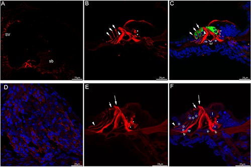
Figure 3.

Acetylated tubulin immunolabeling in the mouse cochlea at P10 and P12. A) An overview of acetylated tubulin (red) labeling in the medial turn of the mouse cochlea at P10. B,C) Detail of acetylated tubulin (red) labeling and myosin VIIa (green) labeling in the organ of Corti in the medial turn of P10 and the merged image +DAPI. Nuel’s space (arrowhead) opened. The neural plexus (asterisks) beneath the IHCs showed strong acetylated tubulin immunolabeling, immunostaining for acetylated tubulin was seen throughout the cell body of inner and outer pillar cells. Noted that in Deiters’ cells, acetylated tubulin expression ran in a longitudinal line, punctuated by small negative areas. Immunolabeling extended from the apical portion of each outer hair cells (arrows) toward the basilar membrane. Positive immunostaining was also detected in the tunnel crossing fibers. D) Detail of acetylated tubulin (red) labeling in the Rosenthal’s canal of the medial turn of P10. Immunostaining of the auditory nerve fibers was maintained. Cell nuclei were counterstained with DAPI (blue). E,F) Detail of acetylated tubulin (red) labeling in the organ of Corti in the medial turn of P12. The labeling for acetylated tubulin was intense and uniform along the full length of the inner and outer pillar cells, its expression in the inner pillar cell phalangeal processes (large arrow) was pronounced. There was moderate labeling in the apical region of the outer pillar cell, the beam bundle (small arrow) interdigitating between the outer hair cells showed acetylated tubulin immunolabeling. Noted that the foot plates of both cell types showed strong labeling. Linear labeling was uniform along most length of Deiters’ cells and was present mainly in the Deiters’ cell bodies (arrowhead), which spanned the distance between the base of the outer hair cells and the basilar membrane. Strong immunofluorescence was clustered in the basolateral region of the inner hair cells (asterisks). Merged image with cell nuclei stained with DAPI (blue). tcf, tunnel crossing fibers; fp, footplate; sb, spiral limbus; D, Deiters’ cell; I, inner hair cell; O, outer hair cell; op, outer pillar cell; ip, inner pillar cell; SV, stria vascularis.
