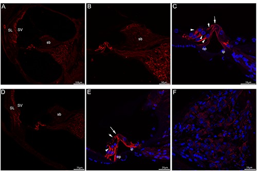
Figure 5.

Acetylated tubulin immunolabeling in the mouse cochlea at P21 and P30. A) A low-magnification view of acetylated tubulin (red) labeling in the medial turn at P21. B) A high-magnification image of acetylated tubulin immunolabeling in the medial turn at P21. C) A higher-magnification image of acetylated tubulin immunolabeling in the organ of Corti in the medial turn at P21. Acetylated tubulin immunolabeling was prominent and uniform in both bundles in each of the pillar cells, the transcellular bundle (large arrow) and beam bundle (small arrow) showed positive immunostaining. Noted that acetylated tubulin immunolabeling in three rows of Deiters’ cells was found at their cup-shaped middle regions (arrowheads). Merged image with cell nuclei stained with DAPI (blue). D) A low-magnification view of acetylated tubulin (red) labeling in the medial turn at P30. E) A higher-magnification image of acetylated tubulin immunolabeling in the organ of Corti in the medial turn at P30. Noted that acetylated tubulin continued to be expressed in Deiters’ cups (arrowheads). Labeling appeared also in both transcellular bundle (large arrow) and the beam bundle (small arrow). Merged image with cell nuclei stained with DAPI (blue). F) Detail of acetylated tubulin (red) labeling in the Rosenthal’s canal of the medial turn of P30. Acetylated tubulin showed no apparent labeling in the somata of spiral ganglion neurons. Cell nuclei were counterstained with DAPI (blue). SL, spiral ligament; SV, stria vascularis; sb, spiral limbus; op, outer pillar cell; ip, inner pillar cell.
