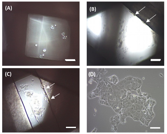
Figure 5.
Optical microscope images of MCF-7 cells seeded on the scaffolds. In 90° scaffolds, cells were attached at the bottom of the well (A). In 45° and 60° scaffolds, cells were attached both on the scaffold and at the well (B,C, respectively). (D) MCF-7 cells in 2D culture. White arrows on the images indicate cells adhered to PCL filaments. Scale bars represent 100 µm.

