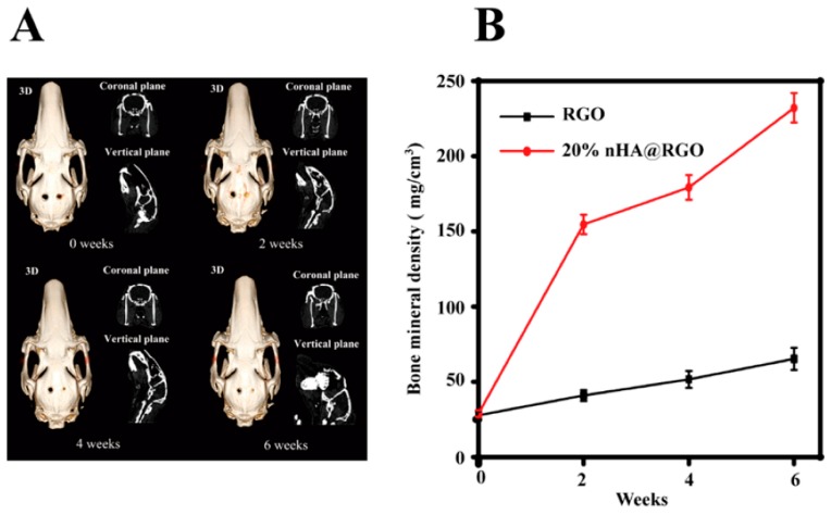
Figure 4.
Proliferation and of bone mineral density (BMD) of rat bone mesenchymal cells. (A) Computed tomography analysis of the defect repair at different time interval. The left defect planted by free reduced graphene oxide scaffold, and the right treated with 20% nHA@RGO scaffold. (B) The change of bone mineral density (BMD) after the scaffold implantation [71] (copyright 2017, Carbon).

