Figure 5.
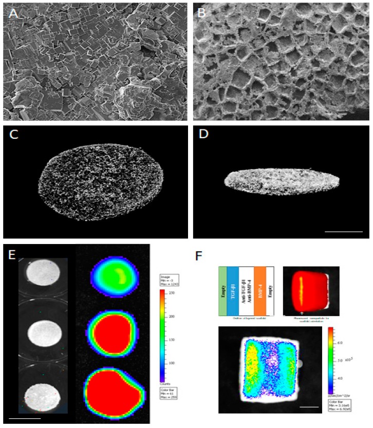
PLGA can be fabricated into scaffolds using gas foaming technique with a water-soluble porogen, salt particles in this case. Scanning electron microscope shows polymer–salt mix after foaming (A) and water leaching (B), demonstrating the uniform pore network. The interconnectivity of the scaffold pore network can be assessed with microcomputed tomography, as seen from the top (C) and side (D) Scale bar = 1 cm. These scaffolds can be fabricated with the PLGA microspheres that release specific payloads, as shown here with a morphogen alone, TGF-β1, in a dose-dependent manner (E). These strategies can be combined with multiple morphogens, TGF-β1 and BMP-4, in this case, along with their pathway-specific inhibitor such as a TGF-β inhibitor, SB43152, and BMP inhibitor Dorsomorphin. Scale bar = 3 cm (F) to create spatially-restricted, morphogen fields within scaffolds. Scale bar = 1.5 cm.

