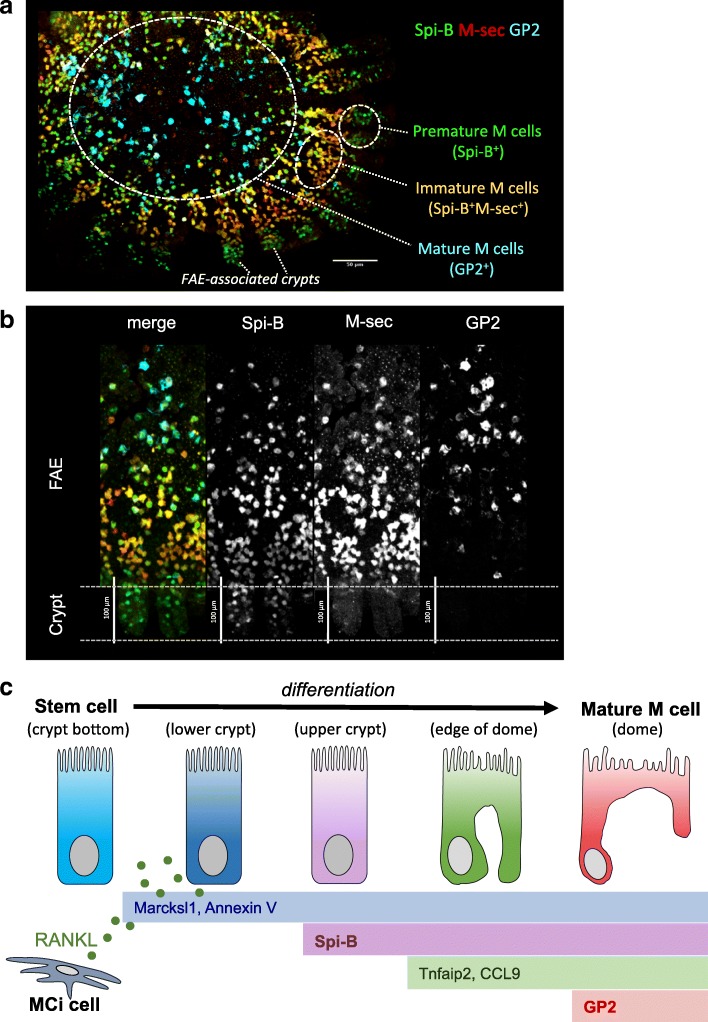
Fig. 2.
Expression of M cell marker molecules during differentiation. a Fluorescence in situ hybridization (FISH) image of Spib, M-Sec, and Gp2 in a mouse FAE specimen is shown. Premature and immature M cells are observed at the crypt and the periphery of dome region, respectively, whereas fully mature M cells are found at the middle and upper dome region. b Enlarged images of the cypt region are shown. c RANKL from stromal M cell inducer (MCi) cells stimulates intestinal stem cells to initiate M cell differentiation. Early (Marksl1, AnnexinV), intermediate (Spi-B, Tnfaip2, CCL9), and mature (GP2) markers are serially expressed during the course of differentiation

