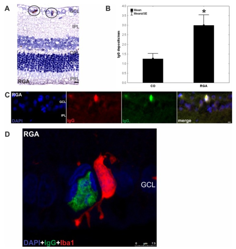
Fig. (2).
IgG deposits and microglia in retina of EAG animals six weeks after immunization with RGC layer homogenate (RGA). A) IgG deposits were observed in retina cross-sections (n = 10 eyes/group) of RGA animals immunized with RGA layer homogenate in an equal volume of incomplete Freund’s adjuvant plus pertussis toxin (circled). B) counts revealed a significantly higher number of these deposits in the RGA group in relation to control animals injected with NaCl in Freund’s adjuvant and pertussis toxin. (p = 0.02). C) DAPI, IgG, and IgG1 staining in a representative retina cross-section of a RGA immunized animal. Co-localization of IgG and IgG1 is evident in the merged rightmost picture. The majority of the detected deposits were of the IgG1 subtype. D) Confocal image of the retina of an RGA animal. A colocalisation of the IgG deposit and an Iba1+ microglia can be seen. Values are mean+/-SE. Abbreviations: GCL = ganglion cell layer, IPL = inner plexiform layer, INL = inner nuclear layer, OPL = outer plexiform layer, ONL = outer nuclear layer, PRL = photoreceptor layer. (*, p<0.05; scale bars in A and C: 10 mm and in D:7.5 mm) (published by S. C. Joachim et al; PLoS One. 2012; 7(7): e40616. doi:10.1371/journal.pone.0040616) [86] Concerning the copy right license: Copyright notice: This is an open-access article distributed under the terms of the Creative Commons Attribution License, which permits unrestricted use, distribution, and reproduction in any medium, provided the original author and source are credited).

