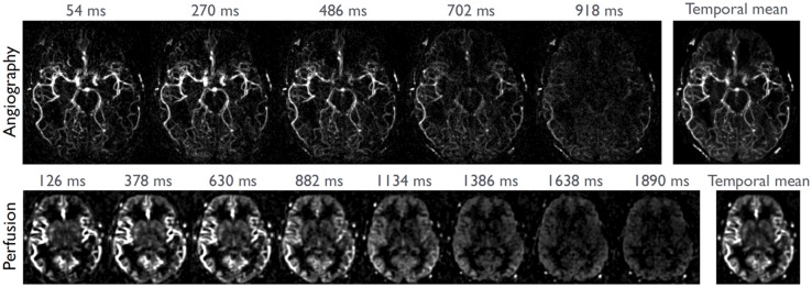
Figure 6.
Combined angiography and perfusion using radial imaging and ASL (CAPRIA) as based upon the Golden Angle Radial approach. Selected angiographic frames are shown from a transverse maximum intensity projection (108 ms temporal resolution, top) as well as a single slice of perfusion data (252 ms temporal resolution, bottom), reconstructed from the same raw data sets. Image courtesy: Thomas Okell (FMRIB Centre, Nuffield Department of Clinical Neurosciences, University of Oxford, Oxford, UK).

