
Figure 2.
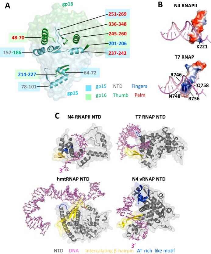
The mechanism of RNAPII heterodimerization and structural organization of the N4 RNAPII promoter recognition motifs. A, surface model of RNAPII-promoter DNA complex with structural elements of gp15 and gp16 subunits that form heterodimerization surfaces overlaid as ribbon models. Subunits and their structural elements are colored as in Fig. 1B. Positions of the elements involved in heterodimerization are colored as corresponding subunits and subdomains in Fig. 1. B, distribution of electrostatic potentials on the surfaces of specificity loops of N4 RNAPII and T7 RNAP. Blue, red, and white colors depict areas with positive, negative, and neutral charges, correspondingly. Residues that form basic patches on the specificity loop tips are shown. C, structural organizations of NTDs in T7-like RNAPs and interaction of their promoter recognition motifs with DNA. NTDs of N4 RNAPII, T7 RNAP, human mitochondrial RNAP, and N4 vRNAP are shown as ribbon models overlaid on their surface models and colored as in Fig. 1. Promoter templates are shown as stick models in magenta.