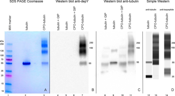
Figure 1.
SDS-PAGE stained with Coomassie Blue (lanes 1–3), Western blotting on PVDF membrane probed with anti-diethoxyphosphotyrosine antibody depY (lanes 4–7), Western blotting probed with anti-tubulin antibody (lanes 8–11), and a capillary electrophoresis Simple Western image (lanes 12–14) probed with anti-tubulin antibody (lanes 12 and 13) or anti-isopeptide antibody (lane 14). All samples were reduced with DTT prior to electrophoresis. The band at 35 kDa for untreated tubulin (lane 12) was an artifact of the Simple Western procedure.

