Figure 3.

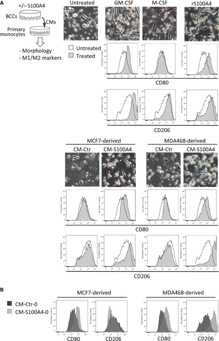
Effect of BCC‐derived CMs on primary human monocytes. (A) The experimental setup is depicted in the cartoon. Primary human monocytes were cultivated for 7 days in the presence of indicated factors: GM‐CSF or M‐CSF (both 50 ng·mL−1), rS100A4 (2 μg·mL−1), CM‐Ctr, and CM‐S100A4 from MCF7 or MDA468 BCCs. Changes in monocyte morphology and expression of M1 and M2 polarization markers, CD80 and CD206, were analyzed by microscopy and flow cytometry, respectively. Representative phase‐contrast images from each condition are shown in the upper panel. Histogram overlays indicate the levels of CD80 and CD206 under each treatment condition (filled histograms) compared to the untreated control (unfilled histograms). (B) Levels of CD80 and CD206 in primary monocytes cultured in CM‐S100A4‐0 (depleted for rS100A4) versus the respective control CM‐Ctr‐0 from MCF7 and MDA468.
