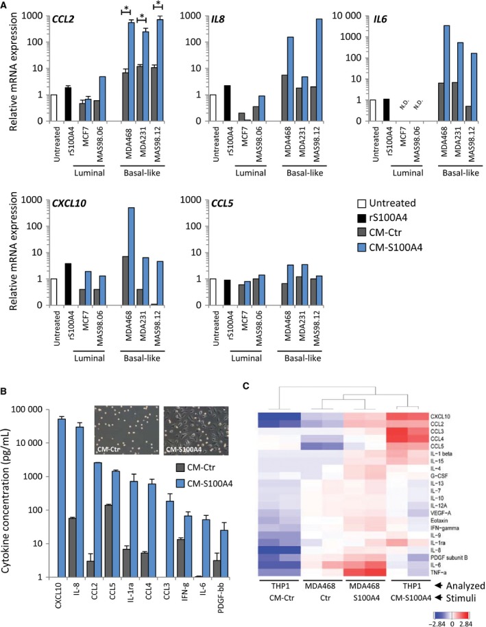
Figure 5.

S100A4‐activated basal‐like BCCs educate THP1 cells to produce cytokines. (A) THP1 cells were cultured for 7 days in CM‐Ctr or CM‐S100A4 from luminal or basal‐like BCCs (‘rS100A4’ – treatment with 2 μg·mL−1 rS100A4 alone) and analyzed for expression of cytokine genes. Relative mRNA expression of indicated cytokines under each condition compared to untreated controls (set to 1). For CCL2, average ± SEM (n = 3, except MAS98.06, where n = 1); the other cytokines were measured in one representative sample from each BCC model. N.D. indicates below the detection limit. (B) THP1 cells were cultured for seven days in CM‐Ctr or CM‐S100A4 from MDA468, washed, and received an ordinary serum‐free medium, and after 3 days, their growth medium was analyzed for cytokines by multiplex immunoassay. The graph indicates the top 10 cytokines elevated in CM‐S100A4 compared to CM‐Ctr. Average concentrations (pg·mL−1) of cytokines from two independent experiments ± SEM. The morphology of the resulting THP1 cells is depicted in the insert. (C) Heatmap with hierarchical clustering of all detectable cytokines (log10‐transformed and median‐centered) in THP1 and MDA468 (Analyzed) in control and S100A4‐stimulated conditions (Stimuli). Two biological parallels for each condition are shown.
