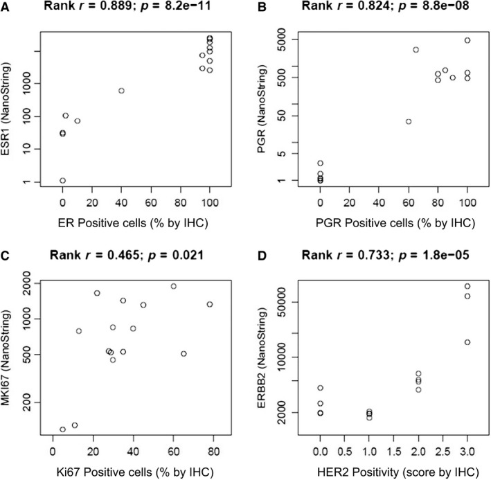
Figure 2.

Comparison between mRNA and IHC analyses. Correlation between mRNA levels and IHC results for (A) ESR1 (ER), (B) PGR, (C) MKI67 (Ki67), and (D) ERBB2 (HER2). mRNA levels were analyzed using NS technology in FNA samples, while IHC was conducted in the corresponding CNB samples. The X‐axes of the scatter plots show the percentage of immune‐positive cells for ER, PGR, and Ki67, and level of HER2 positivity (0 to +3) given by the routine IHC report. The Y‐axes show log‐transformed mRNA expression levels (counts).
