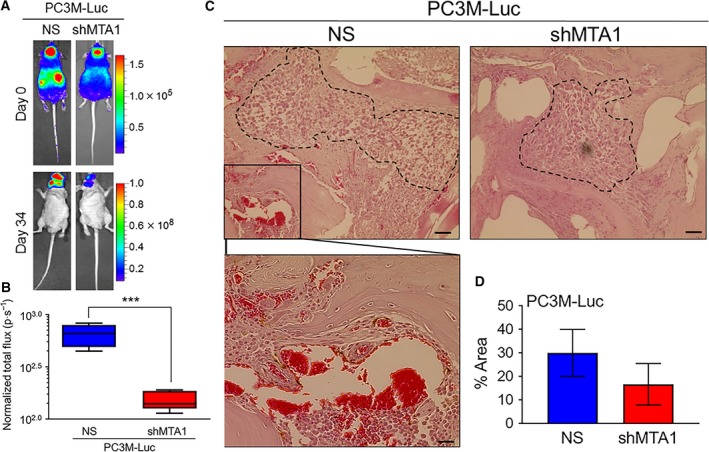
Figure 1.

MTA1 knockdown suppresses formation of bone metastasis in experimental bone metastasis xenografts. (A) PC3M‐Luc‐NS and PC3M‐Luc‐shMTA1 cells were injected into the left ventricle of male athymic mice (n = 5) to colonize the bone. Bioluminescent images of representative tumor metastasis at day 0 (dorsal view) and day 34 (ventral view) are shown. (B) Quantitative analysis of bioluminescent signals of metastasis is shown in normalized total flux (photon per sec). ***P < 0.001 (unpaired t‐test). (C) Representative histology for bone metastatic lesions from two groups of mice is shown. Scale bars, 50 μm. Scale bar (magnified image), 10 μm. (D) Quantitation of the metastatic carcinoma area. Data represent the mean ± SEM of metastatic area from three mice in each group.
