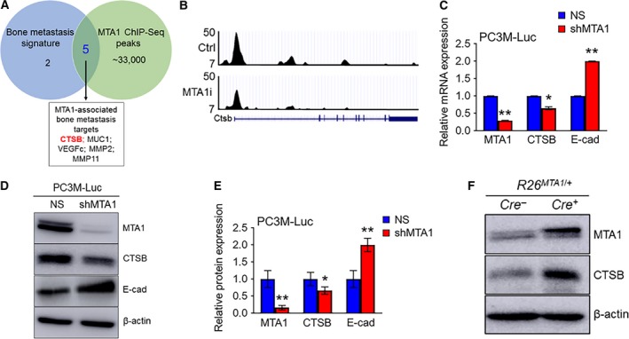
Figure 4.

MTA1 knockdown reduces cathepsin B mRNA and protein levels in PC3M‐Luc prostate cancer cells. (A) Venn diagram depicting a number of genes selected as bone metastatic signature (Kai et al., 2011), number of peaks identified by MTA1 ChIP‐Seq (Dhar et al., 2016), and overlapping MTA1‐associated bone metastasis targets of interest. (B) Comparative analysis of ChIP‐Seq data on MTA1 binding in the prostate tissue of Pten +/− mice expressing high levels of MTA1 (Ctrl, upper panel) and reduced levels of MTA1 (MTA1i, lower panel). Representative MTA1 ChIP‐Seq track for CTSB gene loci at 10‐kb resolution is shown. (C) qRT‐PCR of MTA1, CTSB, and E‐Cad mRNA levels in PC3M‐Luc‐NS and PC3M‐Luc‐shMTA1 cells. (D) Immunoblots of MTA1, CTSB, and E‐cad in PC3M‐Luc‐NS and PC3M‐Luc‐shMTA1 cells. β‐Actin was used as a loading control. (E) Quantitation of immunoblot signals. Data represent the mean ± SEM of three independent experiments. *P < 0.05; **P < 0.01 (two‐way ANOVA). (F) Immunoblots of MTA1 and CTSB in prostate tissues from 13‐week‐old Pb‐Cre +; R26 MTA 1/+ mice and Cre‐negative normal prostate control.
