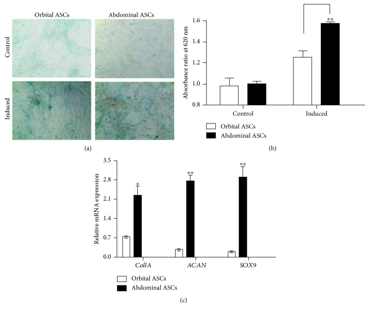
Figure 4.
Chondrogenic differentiation of orbital and abdominal ASCs towards chondrogenesis. (a) Microscopic photograph of ASCs cultured in chondrogenic medium for 21 days and stained with alcian blue. Control cells were grown in DMEM/F12 medium for the same period of time. (b) Quantitation analysis of extracellular matrix production by determining the amount of dye extracted with 6 M guanidine HCl and measured at absorbance 620 nm wavelength. (c) Real-time PCR analysis for genes involved in chondrogenesis, such as Col1A, ACAN, and SOX9. Results are presented as means ± standard error or mean (SEM). ∗p < 0.05 and ∗∗p < 0.01.

