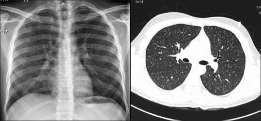
Figure 2.

Composite image of the chest radiograph (left) showing small nodular opacities and high-resolution computed tomography of the thorax (right) showed bilateral profuse evenly distributed micronodules with tree-in-budding; pulmonary artery was normal in size, and there were no associated lymphadenopathy. Expiratory computed tomography did not show any air trapping (not shown)
