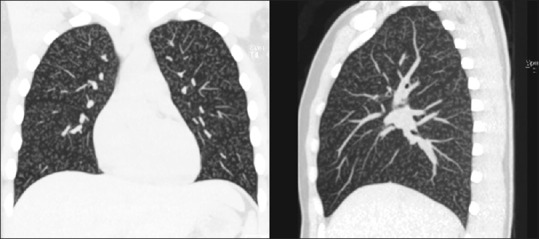
Figure 3.

Coronal and sagittal reconstruction of the maximum intensity projection formatting of the high-resolution computed tomography images of the thorax showing diffuse, extensive, bilateral centrilobular nodules with branching and without any lobar predominance
