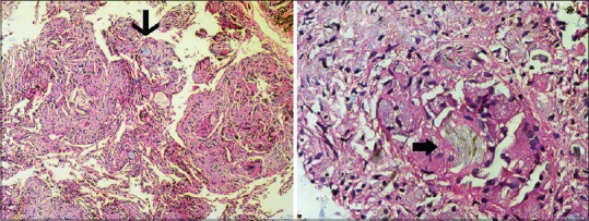
Figure 5.

Photomicrographs of the transbronchial lung tissue biopsy specimen (left) demonstrating perivascular foreign body granulomatous reaction to the talc particles (thin arrow, H and E, ×100). Under polarized light, birefringent 20–95 μm needle-shaped crystals were seen in the granuloma, consistent with talc (block arrow, H and E, ×400)
