
Extended Data Figure 1. DRP1 and MID49 assembly states.
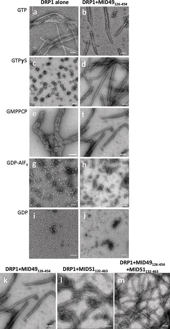
(a-j) DRP1 assembly states visualized with negative stain electron microscopy in the presence of different guanine nucleotides and MID49126-454. Both proteins incubated at 2μM concentration. Bars = 100nm.
(k-m) MID49126-454 and MID51132-463 form indistinguishable assemblies with DRP1. (k) DRP1 plus MID49126-454 and GMPPCP, (l) DRP1 plus MID51132-463 and GMPPCP, (m) DRP1 plus both equimolar MID49 and MID51. Bars = 100nm.