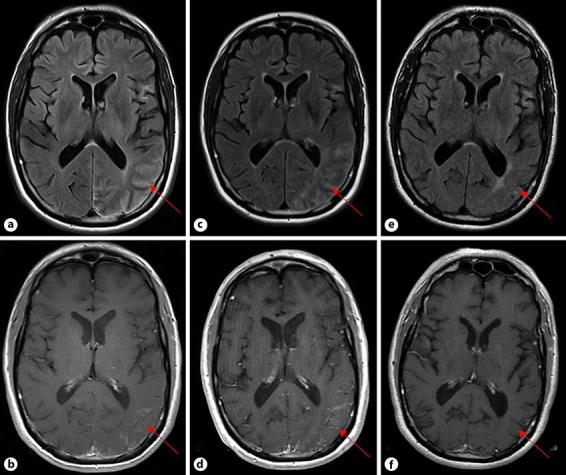
Fig. 1.

Changes on axial MRI over time. a FLAIR image showing signal abnormalities in the left temporal, parietal, and occipital lobes at the time of neurological presentation. b T1+ contrast image demonstrating leptomeningeal enhancement within the left occipital and parietal lobes at the time of neurological presentation. c FLAIR image showing increased signal abnormalities 3.5 months after presentation. d T1+ contrast image demonstrating increased leptomeningeal enhancement 3.5 months after presentation. e FLAIR image showing significant reduction in signal abnormality 22 months after presentation. f T1+ contrast image demonstrating resolution of leptomeningeal enhancement 22 months after presentation.
