Fig. 6.
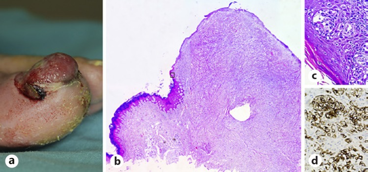
SUM. a A 72-year-old male patient with a 6-month history of rapidly growing erythematous nodular lesion. b Invasive SUM showing the basal and suprabasal layers of the nail matrix and bed epithelium and dermis invaded by an excessive number of melanocytes often displaying severe cellular atypia. H&E, ×4. c In situ SUM displaying nests and lentiginous proliferation of severely atypical melanocytes. H&E, ×40. d Melanocytes are positive for HMB-45. HMB-45, ×40. SUM, subungual melanoma.

