
Figure 2.
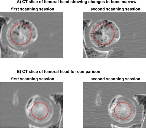
One femur seemed to have lost bone marrow in the femoral head after the first compared to the second scanning session (A). For comparison, we show another femur after the first and the second scanning session (B). The red line depicts the edge of the trabecular ROIs.