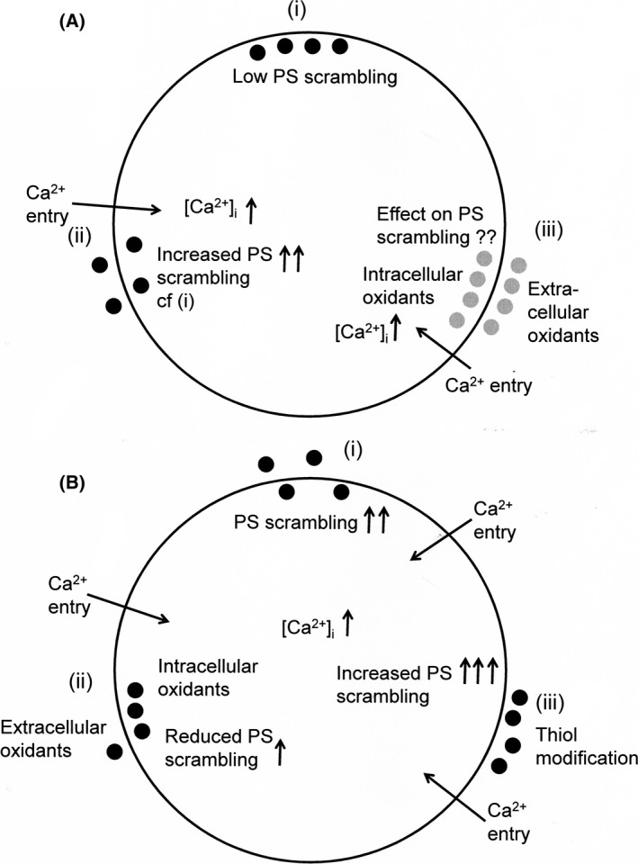
Figure 6.

Schematic digram of stimuli affecting phosphatidylserine (PS) distribution in red cells from patients with sickle cell anaemia. (A) (i) PS is usually confined to the inner leaflet of the lipid bilayer of red cells including sickle cells through high activity of the flippase and low activity of the scramblase, as externalisation is prothrombotic and increases phagocytosis; (ii) elevation of intracellular Ca2+ ([Ca2+]i) via the deoxygenation‐induced cation conductance (or Psickle) or via ionophore promotes PS exposure increasing the possibility of microvascular occlusion; (iii) the effect of oxidants either from within the sickle cell or from the circulation is uncertain. (B) (i) As before, entry of Ca2+ increases PS exposure; (ii) most oxidants (xanthine oxidase/hypoxanthine mixtures, nitrite, phenazine methosulphate) actually reduced Ca2+‐induced PS exposure by about 50% and would reduce thrombosis; (iii) the exception was thiol oxidation which markedly increased externalisation of PS. ●, known PS distribution;  , PS distribution unknown.
