Figure 3.

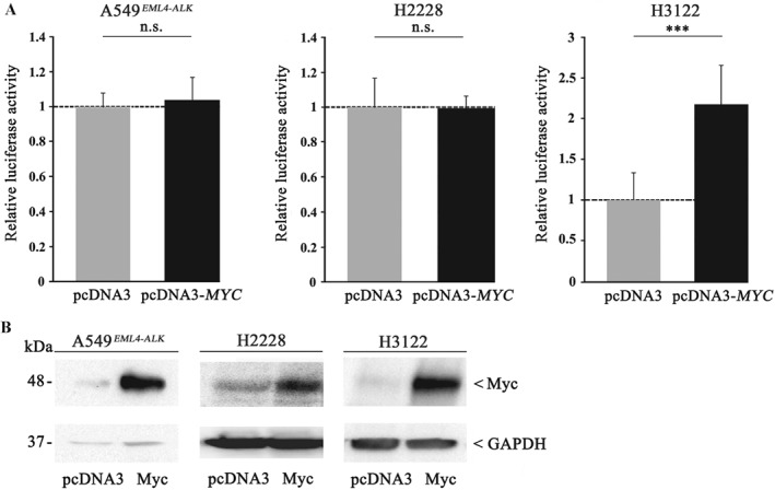
MYC enhances cell proliferation only in TP53 mut but not in TP53 WT ALK+ cell lines. (A) A549EML4–ALK (left), H2228 (middle), and H3122 (right) were transiently transfected with a pcDNA3‐MYC expression plasmid or pcDNA3 vector as control. Twenty‐four hours after transfection, cells were seeded in 96‐well plates and luminescence was measured after 24 h by means of a Centro LB 960 microplate luminometer. Each experiment was performed independently and at least in triplicates, and each measurement was carried out with eight technical replicates. All values are expressed as means ±standard deviation. Statistical significance was evaluated using Student's t‐test: n.s. = not significant, p > 0.05; ***p ≤ 0.001. (B) MYC overexpression was confirmed by immunoblot analysis in A549EML4–ALK (left), H2228 (middle), and H3122 (right) using a polyclonal anti‐MYC antibody. Equal protein loading was ensured by determining GAPDH levels.
