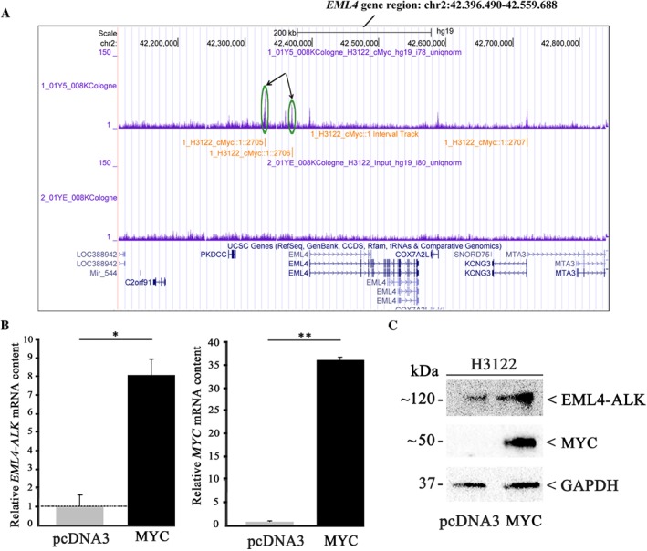
Figure 4.

MYC upregulates EML4–ALK expression in TP53‐mutated H3122 cells. H3122 cells were transiently transfected with a MYC expression plasmid or pcDNA3 vector as control. (A) ChIP‐Seq analyses revealed MYC binding sites within the EML4–ALK promoter region (green circles and arrows). (B) MYC and EML4–ALK mRNA levels were determined by TaqMan gene expression assay. The values shown were normalised to control transfection. All values are expressed as means ±standard deviation. Statistical significance was evaluated using the Student's t‐test: *p ≤ 0.05, **p ≤ 0.01. (C) EML4–ALK and MYC protein amounts were determined by immunoblot analysis using a monoclonal anti‐ALK or a polyclonal anti‐MYC antibody. Equal protein loading was ensured by determining GAPDH levels.
