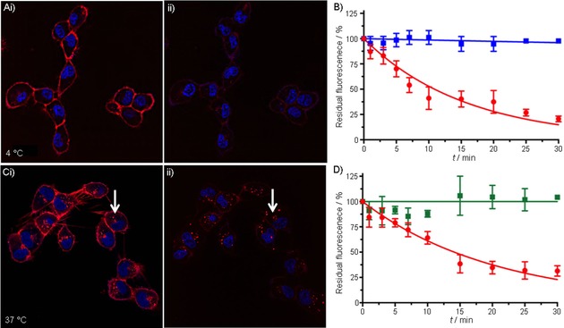
Figure 2.

Confocal‐imaging‐based assessment of membrane availability and internalization kinetics. A) Confocal images of CXCR4‐expressing MDAMB231 X4 cells i: after 1 h incubation with N3‐Cy5‐AcTZ14011 at 4 °C, and ii: 30 min after subsequent addition of Cy7‐DBCO. B) Quantified membrane‐related Cy5‐fluorescence of N3‐Cy5‐AcTZ14011 (•) or non‐azide‐containing Cy5‐AcTZ14011 (▪) over the course of 30 min after addition of Cy7‐DBCO. C) Confocal images of MDAMB231 X4 cells i: after 1 h incubation with N3‐Cy5‐AcTZ14011 at 37 °C, and ii: 30 min after subsequent addition of Cy7‐DBCO (arrows indicate internalized tracer⋅receptor complex located in lysosomes in the cytoplasm). D) Quantified membrane‐related (•) and internalized (▪) N3‐Cy5‐AcTZ14011 fluorescence over the course of 30 min after addition of Cy7‐DBCO. For confocal images: Cy5‐signal in red, nuclear staining in blue.
