-
A
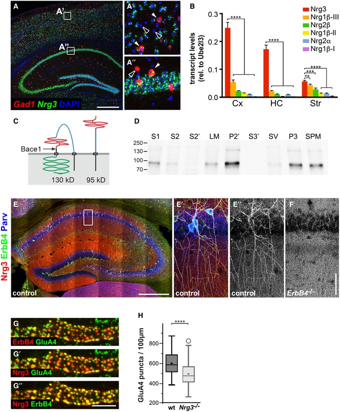
Double fluorescence in situ hybridization using Nrg3 (green)‐ and Gad1 (red)‐specific probes; DAPI was used as a counterstain. Nrg3 is present in Gad1‐positive (filled arrowheads) and Gad1‐negative neurons. (A′) and (A″) show higher magnifications of boxed areas indicated in (A). Nrg3‐negative neurons are indicated by open arrowheads; note that many but not all neurons express Nrg3.
-
B
Comparison of mRNA levels of Nrg3, Nrg1β type III (Nrg1β−III), Nrg2β, Nrg1β type II (Nrg1β−II), Nrg2α, and Nrg1β type I (Nrg1β−I) in the cortex (Cx), hippocampus (HC), and striatum (Str) of juvenile mice (P30–60) by quantitative RT–PCR. Data represent means ± SD of four biological replicates. Two‐way ANOVA with Bonferroni's multiple comparisons test was performed to assess statistical significance (****P < 0.0001, ***P < 0.001, ns = not significant).
-
C
Schematic display of the structure of Nrg3 before (left) and after cleavage by Bace1 (right). The following domains of Nrg3 are indicated: black, N‐terminal intracellular domain and transmembrane domain; blue, extracellular domain; red EGF domain; black, stalk region with Bace1 cleavage site indicated by an arrow and C‐terminal transmembrane domain; and green, intracellular domain.
-
D
Detection of Nrg3 by Western blot analysis in fractions from brain lysates: S1, crude lysate; S2, cytosol and light membranes; S2′, cytosol; LM, light membranes; P2′, crude synaptosomes; S3′, synaptosomal cytoplasm; SV, synaptic vesicles; P3, synaptosomal membranes; and SPM, synaptic plasma membranes.
-
E, F
Immunohistochemical analysis in the hippocampus of wildtype (E, E′, E″) and ErbB4 mutant mice (F) at 2 months of age using Nrg3‐, ErbB4‐, and parvalbumin (Parv)‐specific antibodies. The white box in (E) is displayed magnified in (E′, E″). In control mice, Nrg3 is enriched on dendrites of ErbB4+ PV interneurons, compared to the surrounding neuropil (E′, E″). In ErbB4 mutants, the Nrg3 enrichment on PV interneurons is not apparent (F).
-
G
Nrg3, ErbB4, and GluA4 were identified by immunohistology and co‐localized in synaptic puncta on dendrites of interneurons in the stratum radiatum of the hippocampal CA1. Note that (G) displays Nrg3/ErbB4, (G′) Nrg3/GluA4, and (G″) ErbB4/GluA4 signals of the same triple‐stained image; false colors were assigned for better signal visualization.
-
H
Quantification of GluA4 puncta present on ErbB4+ dendrites in the CA1 stratum radiatum of adult wildtype (wt) and Nrg3
−/− mice (age P90–120). Data are presented as box plots with Tukey's whiskers and outliers; means are indicated by a plus symbol. n = 65 (wt) and n = 62 (Nrg3
−/−) from five animals each. Unpaired t‐test (two‐tailed) with Welch's correction was performed to assess statistical significance (****P < 0.0001).
Data information: Scale bars: 500 μm (A, E), 50 μm (F), and 5 μm (G″).

