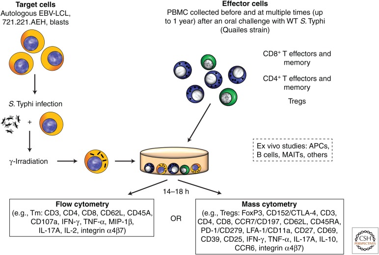
Figure 1.
In vitro stimulation of peripheral blood mononuclear cells (PBMCs) isolated from wild-type (WT) S. Typhi–exposed participants with S. Typhi–infected targets and ex vivo studies: Experimental design. PBMCs obtained before and at various times after oral exposure to the WT S. Typhi Quailes strain were incubated in vitro with autologous Epstein–Barr virus transformed lymphoblastoid B-cell lines (EBV-LCL), 721.221.AEH EBV-LCL (to measure HLA-E-restricted responses) or blasts infected with WT Salmonella enterica serovar Typhi (S. Typhi). After an overnight incubation, cells were fixed and stained for flow cytometry (14-color) or mass cytometry (up to 34 metal-conjugated monoclonal antibodies) depending on whether CD4+ or CD8+ T effector and/or memory subsets (TEM), or regulatory (Tregs) T cells were evaluated. Activation and other characteristics of B cells, antigen-presenting cells (APCs), mucosal-associated invariant T cells (MAITs), or other populations were evaluated ex vivo without stimulation. IFN-γ, Interferon γ; TNF-α, tumor necrosis factor α, MIP-1β, macrophage inflammatory protein-1β, IL-17A, interleukin 17A; FoxP3, forkhead box P3; CTLA-4, cytotoxic T-lymphocyte-associated antigen 4; LFA-1/CD11a, lymphocyte function-associated antigen-1; CD279/PD1, programmed cell death-1; CCR6, C-C chemokine receptor type 6; CCR7, C-C chemokine receptor type 7.

