
Figure 2.
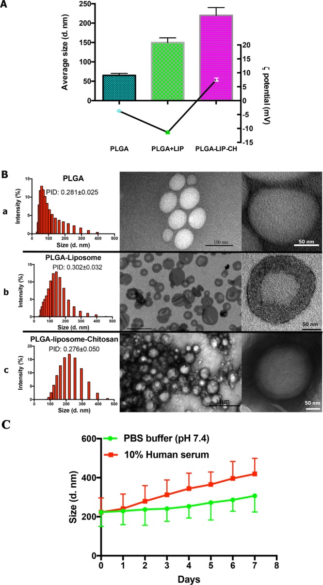
Structure of the multilayered NPs. (A) Hydrodynamic size and ζ potential and (B) morphological properties of MLNPs. TEM images (B, right panels) show the structure of PLGA NPs (panel a), PLGA–liposome NPs (panel b), and Ch-MLNPs (panel c). Dynamic light scattering results with PDI of the NPs at different fabrication stages (B, left panels). (C) Stability of Ch-MLNPs in different buffers.