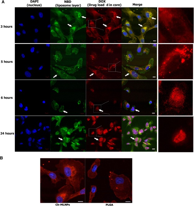
Figure 5.
Confocal microscopic analysis of intracellular trafficking of Ch-MLNPs with DOX loaded in the core in MDA-MB-231 cells. (A) NP distribution at 3, 5, 6, and 24 h post-incubation. Nuclei were labeled with DAPI and liposomes were labeled with NBD (green). DOX, loaded in the core, is self-fluorescent (red). Arrows point to the intracellular tracking of NPs and loaded drugs at different culture times. (B) The intracellular distribution of Ch-MLNPs and PLGA in cancer cells (MDA-MB-231) after incubation for 3 h. Scale bar represents 10 μm.

