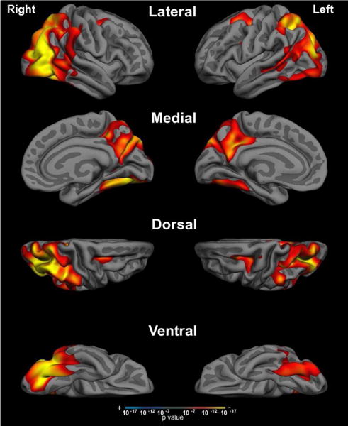
Figure 2. Cortical atrophy in PCA.

Compared to age-matched control participants, whole-brain cortical thickness analysis of PCA patients revealed significant bilateral occipital, lateral temporal, and parietal cortical atrophy with sparing of the medial temporal and frontal lobes. Threshold set at p <10−7.
