
Figure 2. Gao-binge decreases the number of lysosomes with increased accumulation of autophagosomes and inhibits TFEB in mouse livers.
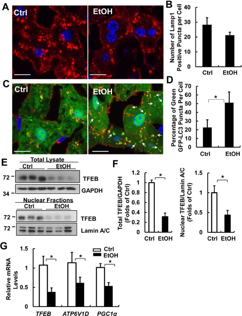
Male GFP-LC3 transgenic mice were treated with Gao-binge model and cryosections of mouse livers were subjected to immunostaining for Lamp1 followed by confocal microscopy. Representative images of immunostaining of Lamp1 (A) and quantified number of Lamp1 positive vesicles are shown (B). Scale Bar: 10 μm. Data are means ± SE (n=3-4). More than 50 cells were counted in each mouse. (C) Representative images of the colocalization of Lamp1 with GFP-LC3 puncta are shown. Arrows denote the green-only GFP-LC3 puncta. (D) Percentage of GFP-LC3 puncta that are not colocalized with Lamp1 positive vesicles. Data are means ± SE (n=3-4). More than 50 cells were counted in each mouse. *p<0.05; Student t test. (E) Male C57BL/6J WT mice were treated with Gao-binge model. Total lysates and nuclear fractions from mouse livers were subjected to western blot analysis. (F) Densitometry analysis of (E). (G) mRNA from mouse livers was used for qPCR. Results were normalized to 18s and expressed as fold change compared to Ctrl group. Data shown are means ± SE (n =4). *p<0.05; Student t test.