
Figure 4. Gao-binge activates mTOR in mouse livers.
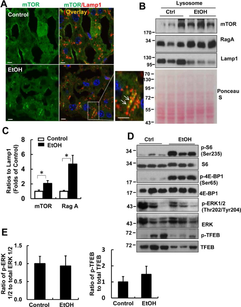
Male C57BL/6J WT mice were treated with Gao-binge model. (A) Cryosections of mouse livers were subjected to immunostaining for mTOR (green) and Lamp1 (red) followed by confocal microscopy. Representative images are shown. Right panel is an enlarged photograph from the boxed area. Arrows denote the colocalization of mTOR with Lamp1. Scale Bar: 10 μm. (B) Lysosomal fractions from mouse livers were subjected to western blot analysis. Membrane was pre-stained with Ponceau S as a loading control. (C) Densitometry analysis of (B) are shown (means ± SE, n=3). *p<0.05; Student t test. (D) Total liver lysates were subjected to western blot analysis. (E) Densitometry analysis of (D) are shown (means ± SE, n=3).