
Fig 3.
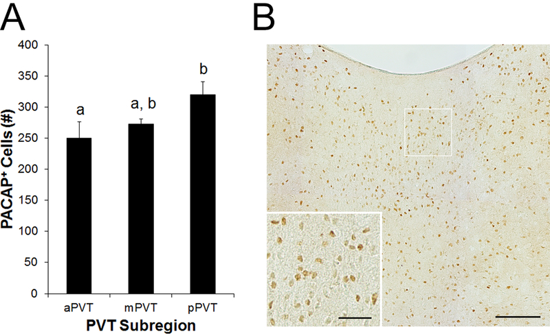
Cells expressing pituitary adenylate cyclase-activating polypeptide (PACAP) protein in the paraventricular nucleus of the thalamus (PVT) of Sprague-Dawley rats (N = 5), as assessed using immunohistochemistry with an antibody that targets both PACAP isoforms (Experiment 2). A. Quantification of cells expressing PACAP in the anterior PVT (aPVT), middle PVT (mPVT), and posterior PVT (pPVT). Bars labeled with the same letter are not significantly different from each other. Values are mean ± SEM. B. Photomicrograph showing PACAP in the mPVT. Inset is a higher magnification of the image marked with a white square. Scale bars = 100 μm in main image and 30 μm in inset.