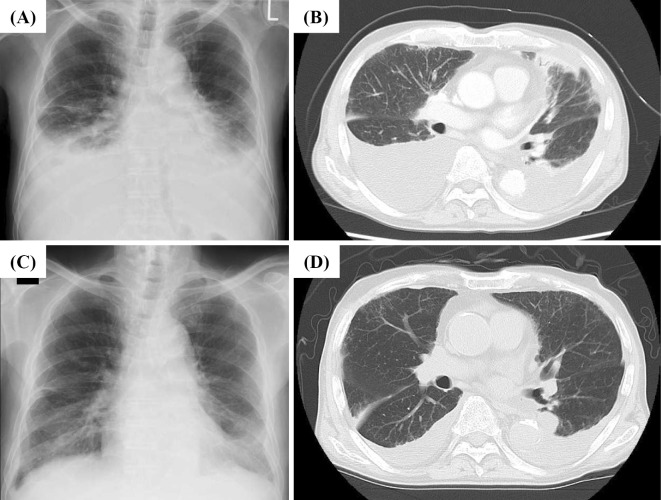
Figure 1.
Changes in the level of pleural effusion after treatment. A chest X-ray (A) and contrast-enhanced CT scan (B) on admission day showed massive bilateral pleural effusion. Contrast-enhanced chest CT of the neck to pelvis also demonstrated subpleural ground-glass opacity in both lungs, consolidation of the left lobe and mild mediastinal lymphadenopathy. No mass lesions or pleural thickening were found. After 4 weeks of treatment with systemic steroids, a chest X-ray (C) and CT scan (D) showed markedly decreased levels of bilateral pleural effusion.

