Figure 2.
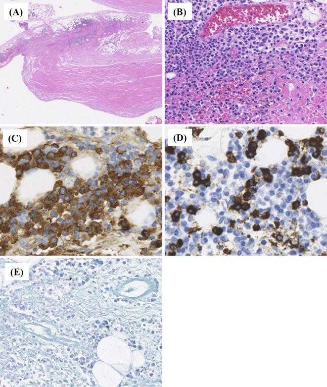
Histopathological evaluation of the pleura and pleural effusion. A pleural biopsy showed the presence of dense lymphoplasmacytic infiltrate [(A): Hematoxylin and Eosin (H&E) staining, ×40; (B): H&E staining, ×100]. Immunohistochemically, many IgG4-poisitive plasma cells are identified (IgG4-positive plasma cells >50/HPF, IgG4+/IgG+cell ration <40%). [(C): immunohistochemical staining for IgG, ×400; (D): immunohistochemical staining for IgG4, ×400]. (E) Ziehl-Neelsen staining identified no acid-fast bacteria.

