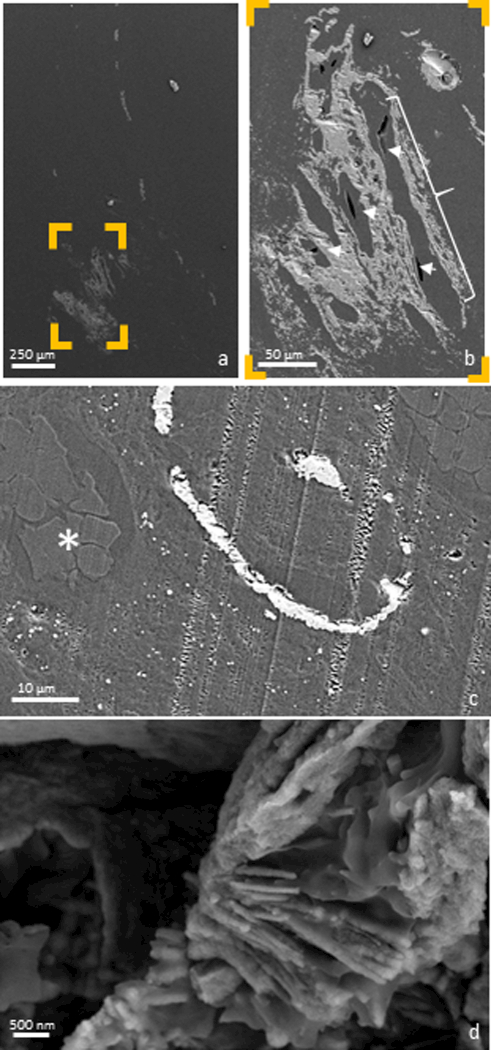
Figure 2. Examining the distal papilla:

Anatomically-specific minerals were observed by imaging the surface of an ultrasectioned distal papilla using an electron microscope. A mineralized tubule can be seen coursing distally down the entire length of the papilla in (a), with nearby mineralized interstitium shown in detail (b). Tubular lumens were patent (b, arrow heads), despite their surrounding tissues being mineralized (b, bracket). Further interstitial mineral is identified in (c), where intravascular erythrocytes (*) were observed in the adjacent mineralized tubular wall. A mineralized collagen fibril, indicative of interstitial (as opposed to intratubular) mineralization was also identified (d).
