Figure 3.
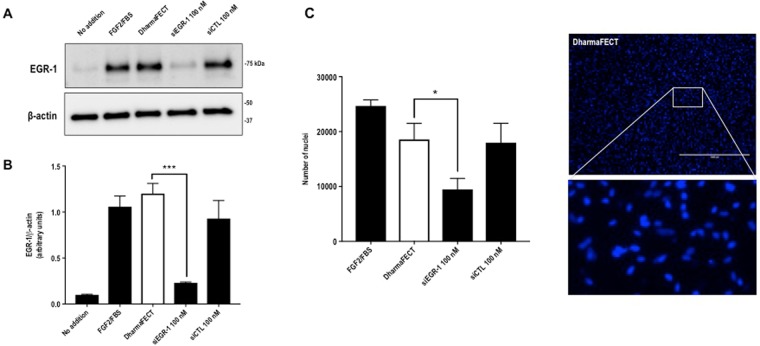
EGR-1 mediates migration in SMC exposed to FGF2. (A) SMC rendered growth quiescent in Waymouth’s medium containing 0.05% FBS were treated with 100 nm siRNA, siCTL or DharmaFECT then incubated with 50 ng/ml FGF2 for 1 h. Western bloting was performed with total cell lysates. Each blot is representative of 2 independent experiments. (B) Band intensity from 3 independent experiments was quantified using NIH Image J and the EGR-1 intensity expressed as a proportion of β-actin intensity per experiment. Two of the three blots were imaged under identical parameters on a LAS 4000 imager. Error bars represent SEM. Statistical significance was assessed by one-way ANOVA. (C) SMC in Waymouth’s containing 20% FBS were seeded into 24-well plates fitted with 0.8 µm Transwell inserts. After 48 h, the medium was changed to Waymouth’s containing 5% FBS and the cells were incubated for 48 h. siRNA, siCTL or DharmaFECT alone was added to the upper chamber at 100 nM and ratio of 1:2 in Waymouth’s medium containing 5% FBS without antibiotics. The medium in the lower chamber was changed to Waymouth’s containing 50 ng/ml FGF2 in 5% FBS. The cells were left for 48 h. Nuclei were quantified using NIH Image J software. Data represents the mean ± SEM of the means of 4 independent experiments. Statistical significance was assessed by one-way ANOVA. *P < 0.05, ***P = 0.0001. A representative image of DAPI stained nuclei from the DharmaFECT group and an enlargement are shown. The scale bar represents 1000 µm.

