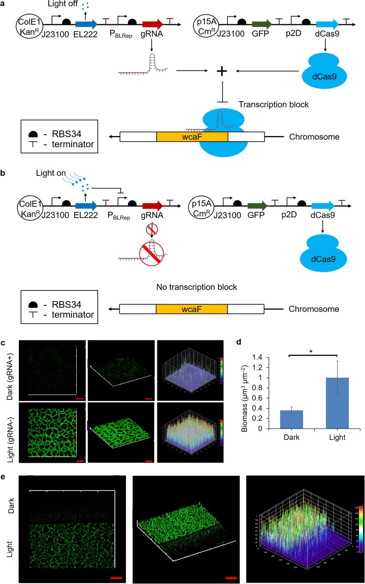
Figure 3.
Using blue light to regulate wcaF gene expression can control biofilm thickness spatially. (a) Gene circuits that consist of constitutive dCas9 expression plasmid and blue light repressible gRNA expression plasmid. gRNA would be expressed and hence wcaF gene would be repressed in dark condition. (b) In contrast, the gRNA would be repressed and wcaF gene would be expressed when exposed to blue light. (c) Control biofilm thickness in spatial. Even the biofilm was cultured on the same microscope slide in the same compartment of medium, confocal images show that only the area that exposed to blue light was able to form thick biofilm. (d) The biomass on the region which was exposed to light was around 3 folds more than that of dark region. (e) At the light-dark boundary, the biofilm thickness showed difference at each side. The three-dimensional plot of GFP intensity also shows the biofilm thickness difference in spatial at the light-dark boundary. Scale bars, 50 μm. All data are represented as mean ± std_dev. *indicates P value < 0.05.

