Figure 3.
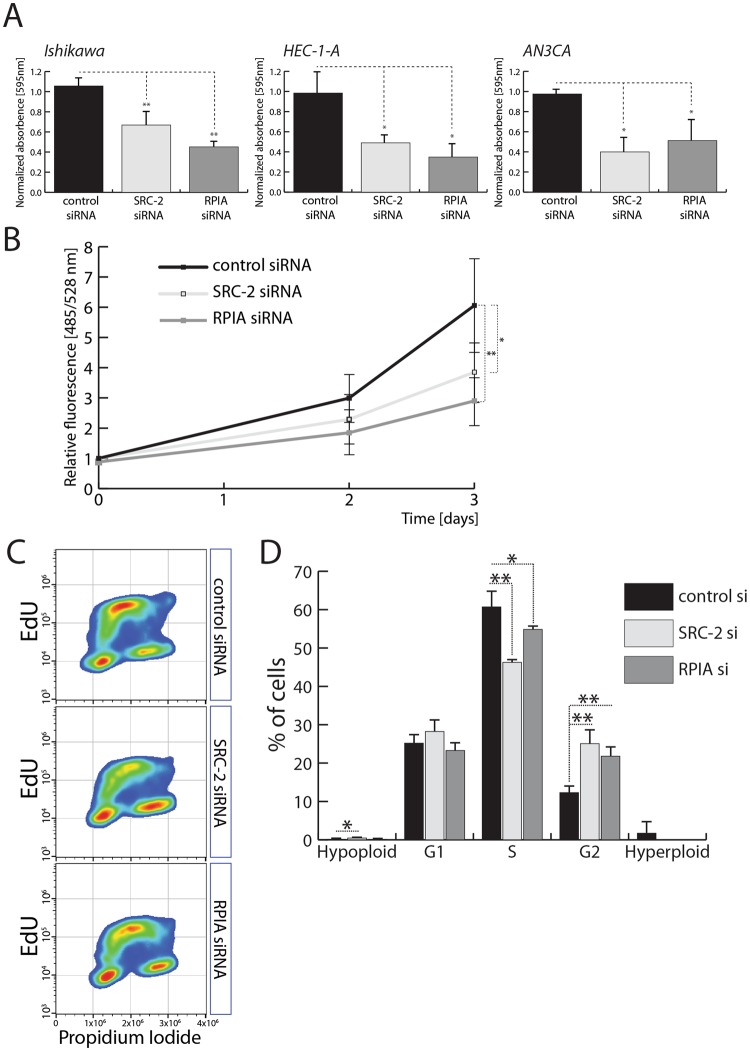
Expression of RPIA is required for EC cell proliferation. (A) Quantification of colony formation assays performed in EC cell lines – Ishikawa, HEC-1-A, and AN3CA – after control, SRC-2, and RPIA siRNA-mediated knockdown. (B) DNA content-based proliferation assay performed with Ishikawa cells after control, SRC-2, and RPIA downregulation. (C) Examples of scatterplots representing measurements of propidium iodide and EdU incorporation of Ishikawa cells transfected with control, SRC-2, and RPIA siRNAs. (D) Quantification of hypo- and hyperploid cell populations and cells in G1, S, and G2 phases of the cell cycle evaluated by quantification of PI and EdU intensity as presented in (D).

