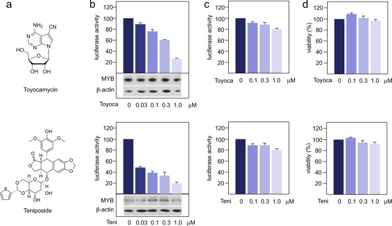
Figure 3.
Myb-inhibitory activity of selected compounds. The figure shows the chemical structures of toyocamycin and teniposide (a), the luciferase activity of Hek-Myb-Luc cells (b) or Hek-Luc cells (c) after treatment for 12 hours with the respective compound and the impact of each compound on the viability of the Hek-Myb-Luc cells (d). The bottom panels in b show the expression of MYB and β-actin. Columns and standard deviations for luciferase activity are based on three independent experiments with two replicate samples in each case. Viability assays are based on two independent experiments with six replicate samples in each case.

