. 2018 Aug 30;27(4):309–319. doi: 10.5607/en.2018.27.4.309

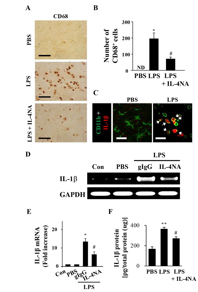
Fig. 4. Neutralization of IL-4 downregulates IL-1β expression in the SN in vivo. Animals were intranigrally infused with LPS (5 µg/3 µl) or PBS as a control in the presence or absence of gIgG or IL-4NA and sacrificed 1 d later for immunohistochemical analysis (A~C), RT-PCR (D, E), and ELISA (F) of the SN. Photomicrographs (A) and the quantification (B) of CD68+ cells in the SN. (C) Colocalization of IL-1b (red) within CD11b+ cells (green). Arrows indicate merged cells. Representative photograph (D) and histogram for the quantification (E) of RT-PCR shows that IL-4NA decrease mRNA expression of IL-1β in the SN. (F) The amounts of IL-1b in the SN were measured using a ELISA technique. Data are presented as the means±SEM of 3 to 5 animals per group. *p<0.01, **p<0.001, compared with PBS; #p<0.01, compared with LPS or LPS with gIgG (ANOVA and Student-Newman-Keuls analysis). Scale bars: A, C: 50 µm.

Fig. 4