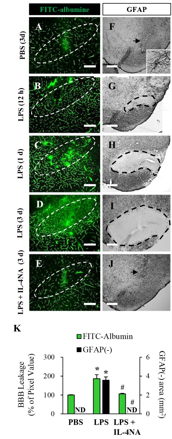
Fig. 5. Neutralization of IL-4 prevents disruption of BBB and astrocytes in the SN in vivo. Animals were administered LPS or PBS in the presence or absence of gIgG or IL-4NA and sacrificed at indicated time point. (A~E) Focal leakage of the BBB was demonstrated by FITC-albumin microangiography. The dashed lines indicate the SNpc. (F~J) The SN tissues were immunostained with anti-GFAP antibody for astrocyte. Dotted lines indicate the lacking area of GFAP staining in the SN. Arrows indicate syringe track. (K) Quantification of BBB leakage density and GFAP immuno negative (i.n.) area at 3 day. Data are presented as the means±SEM of Three to five animals per group. *p<0.01, significantly different from PBS; #p<0.01, significantly different from LPS or LPS with gIgG (ANOVA and Student-Newman-Keuls analysis). Scale bars: A~E, 500 µm; F~J, 200 µm.

