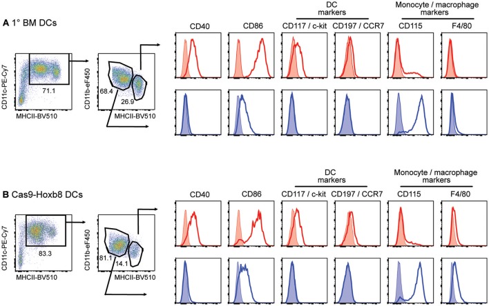
Figure 5.
Phenotype of primary bone-marrow derived DCs (1° BM DCs) and Cas9-Hoxb8 DCs in CM-CSF cultures before LPS activation. Flow cytometric analysis of cells cultured for 8 days in the presence of GM-CSF. Without LPS activation, both (A) 1° BM and (B) Cas9-Hoxb8 cells were subdivided on the basis of CD11c, MHCII and CD11b expression. Histograms show surface expression of the markers indicated on MHCIIhiCD11blo (red) and MCHIIintCD11bhi (blue) myeloid cell subsets. Data are representative for three independent experiments.

