
Figure 2.
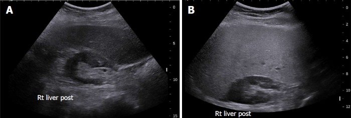
Grey-scale ultrasound in non-alcoholic fatty liver disease. A: 47-year-old female with increased echogenicity of the liver relative to the right kidney, a classic sonographic finding of hepatic steatosis. The patient had elevated serum liver enzymes and underwent a liver biopsy for NASH evaluation; B: 51-year-old female who underwent liver biopsy as part of clinical follow-up. On evaluation of liver pathology, there was no steatosis. Ultrasound image shows normal echogenicity of the liver parenchyma, which is only slightly hyperechoic relative to the renal parenchyma.