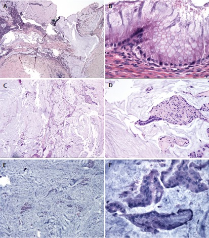
Figure 2.

Histological classification of pseudomyxoma peritonei[25]. This figure highlights some of the characteristic findings in the different histological types of pseudomyxoma peritonei. A, B: Disseminated peritoneal adenomucinous (DPAM) is demonstrated in (A and B) with paucicellular mucin pools (A) and scant strips of low-grade neoplastic epithelium (B); C, D: Peritoneal mucinous carcinamatosis-intermediate (PMCA-I) is demonstrated in (C and D). PMCA-I is less cellular than PMCA, but the degree of atypia exceeds that of DPAM (D); E, F: Highlight PMCA, with mucin cells with abundant epithelium (E) and malignant cytological features (F).
