
Figure 3.
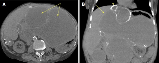
Computed tomography of pseudomyxoma peritonei[26]. This is an axial computed tomography scan. A: Cystic accumulations of mucus (arrows) surrounded by calcified rims; B: A coronal reconstruction representing cystic accumulations in the upper abdomen and the liver.