
Figure 4.
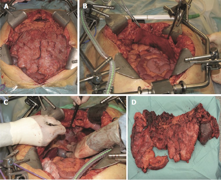
Intraoperative pictures of cytoreductive surgery[11]. This is a figure depicting various stages of cytoreductive surgery. A: A view of a pseudomyxoma patient’s abdominal cavity immediately after laparotomy; B: Stripping of the right anterior peritoneum; C: Depicts stripping of the right subphrenic peritoneum; D: An image of the resected terminal ileum, colon, and spleen affected by pseudomyxoma peritonei.