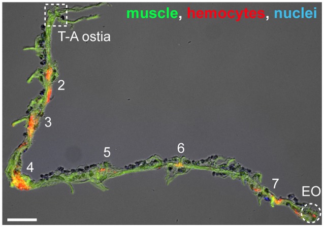
Figure 4.

Hemocytes on a resected heart. Image of a resected heart where muscle (phalloidin; green), hemocytes (CM-DiI; red), and nuclei (Hoechst 33342; blue) have been labeled. In this specimen there are no hemocytes at the thoraco-abdominal ostia (T-A ostia), but there are hemocytes at the periostial regions (abdominal segments are numbered) and at the posterior excurrent openings (EO). Scale bar: 100 μm.
